August 20, 2021
New Imaging Tool Visualizes Cell Functions in a Microphysiological System (TI-FRIS Fellow Dr. Yuji Nashimoto)
A microphysiological system (MPS), also known as an organ-on-a-chip, is a 3D organ construct using human cells that help reveal how organs respond to drugs and environmental stimuli.
Now, a group of researchers led by TI-FRIS Fellow Dr. Yuji Nashimoto and his mentor Prof. Hitoshi Shiku at Tohoku University has developed a new analytical method that visualizes cell functions in MPS using scanning probe microscopy (SPM).
SPM differs from optical microscopy since it employs fine probe scanning over a sample surface and then exploits the local interactions between the probe and the surface. The biggest advantage of SPM over conventional microscopy is that physical and chemical conditions can be acquired rapidly and as a high-resolution image.
In this study, SPMs evaluated a vascular model (vasculature-on-a-chip) by scanning electrochemical microscopy (SECM) and scanning ion conductance microscopy (SICM). Using these SPMs, the researchers quantified the permeability and topographical information of the vasculature-on-a-chip.
"MPS shows potential to recapitulate the physiology and functions of their counterparts in the human body. Most research on this topic has focused on the construction of biomimetic organ models. Today, there is an increasing interest in developing sensing systems for MPS" said Dr. Nashimoto at Tohoku University.
Some have touted electrochemical sensors to monitor MPS. However, most electrochemical sensors cannot acquire the spatial information of cell functions in MPS because they have only one sensor per one analyte. In contrast, SPM provides spatial information about cell functions rapidly.
"Our research group has developed various electrochemical imaging tools, SPMs and electrochemical arrays," explained Prof. Shiku.
This research was published in Advanced Healthcare Materials on August 20, 2021, and a press release was issued from Tohoku University.
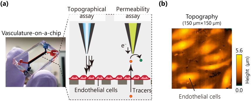
Endothelial functions (permeability and topography) in a microphysiological system (MPS) are electrochemically visualized using scanning probe microscopies (SPMs). The analytical system is a new means to evaluate MPS or organ-on-a-chip (OoC).
Publication Details:
Yuji Nashimoto, Minori Abe, Ryota Fujii, Noriko Taira, Hiroki Ida, Yasufumi Takahashi, Kosuke Ino, Javier Ramon Azcon, Hitoshi Shiku
Advanced Healthcare Materials
"Topography and Permeability Analyses of Vasculature-on-a-Chip using Scanning Probe Microscopies"
DOI: 10.1002/adhm.202101186
https://doi.org/10.1002/adhm.202101186
Press Release:
Tohoku University
FRIS, Tohoku University
https://www.fris.tohoku.ac.jp/en/feature/topics/detail---id-972.html
https://www.fris.tohoku.ac.jp/en/feature/topics/detail---id-972.html